
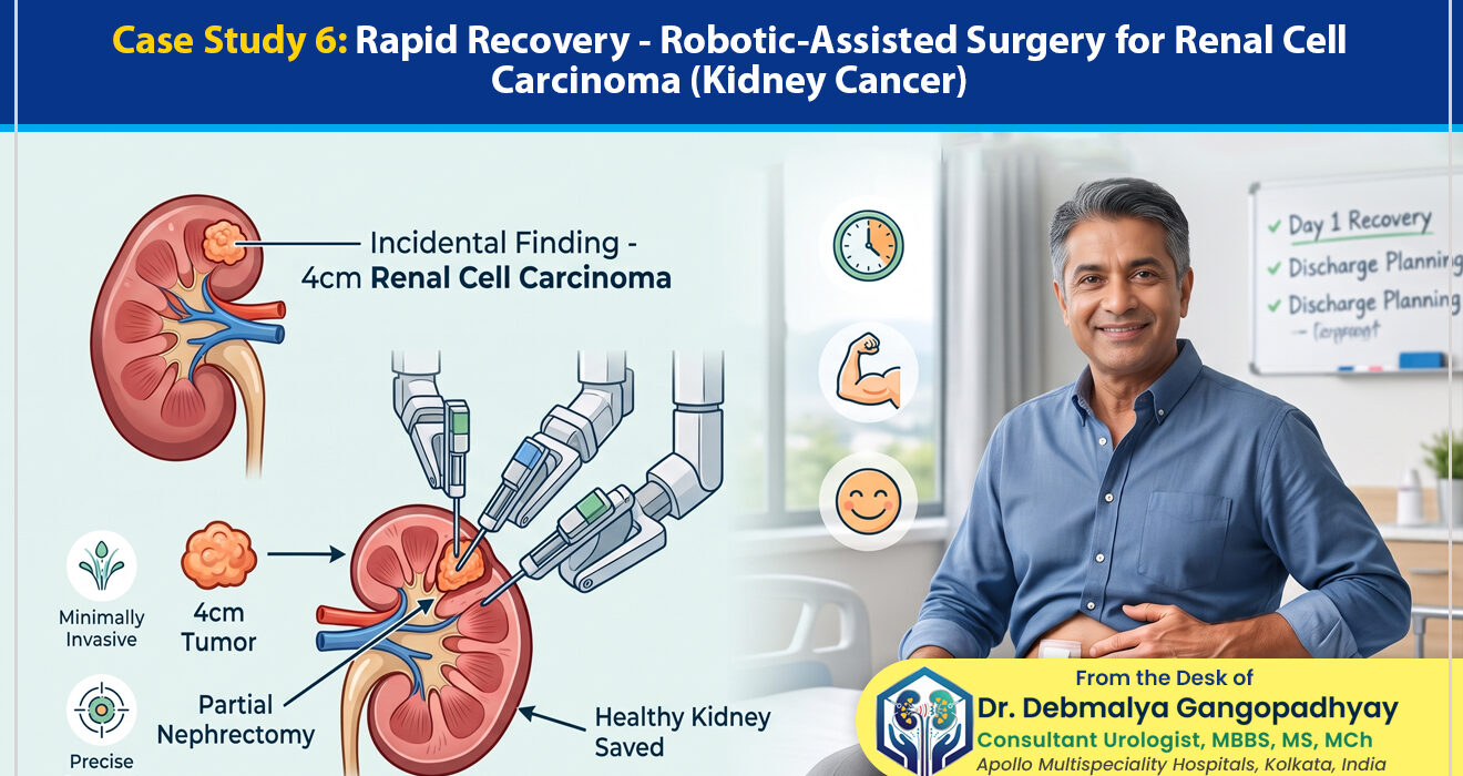
Case Study 6: Rapid Recovery – Robotic-Assisted Surgery for Renal Cell Carcinoma (Kidney Cancer)
Table of Contents
Toggle‘Rapid Recovery – Robotic-Assisted Surgery for Renal Cell Carcinoma (Kidney Cancer)’ – from the desk of Dr. Debmalya Gangopadhyay, Senior Consultant Urologist, Apollo Multispeciality Hospitals, Kolkata, India
Hearing the word “cancer” is one of the most terrifying experiences a human being can endure. Time seems to stop, the room spins, and a flood of questions and fears rush into the mind. When the diagnosis is Kidney Cancer—medically known as Renal Cell Carcinoma (RCC)—patients often immediately envision the worst: the complete loss of their kidney, massive open surgeries, agonizing months of recovery, and the looming dread of dialysis.
However, we are living in a golden era of surgical innovation. The narrative around kidney cancer has drastically changed over the last decade. Today, a kidney cancer diagnosis does not automatically mean losing the entire organ, nor does it guarantee a painful, prolonged hospital stay.
In this comprehensive, step-by-step case study, we will walk you through the remarkable journey of a middle-aged man whose life was turned upside down by a silent, hidden kidney tumor. We will explore how modern medicine, specifically through Robotic Urology Surgery in Kolkata, allowed us to surgically remove the cancer while saving his healthy kidney, leading to a remarkably rapid recovery.
By breaking down the Patient Background and Initial Assessment, the Diagnostic Process and Confirmation, the Line of Treatment, the meticulous Follow-Up, and the ultimate Success story, we aim to humanize this medical journey. If you or a loved one are facing a similar diagnosis, this guide will serve as a beacon of hope, demystifying the process and proving that there is life—a highly active, normal life—after kidney cancer.
Part 1: Understanding the Silent Invader – Renal Cell Carcinoma (RCC)
Before we introduce our patient, it is crucial to understand what Renal Cell Carcinoma is and why it behaves the way it does.
The kidneys are two bean-shaped organs, each about the size of a fist, located just below the rib cage on each side of your spine. They are the body’s master filtration system, removing waste products and excess fluid from the blood to produce urine. They also play a vital role in regulating blood pressure and stimulating the production of red blood cells.
Renal Cell Carcinoma (RCC) is the most common type of kidney cancer in adults. It begins in the lining of very small tubes (tubules) within the kidney.
The Phenomenon of the “Incidentaloma”
In the past, kidney cancer was usually diagnosed only when it had grown large enough to cause obvious symptoms: blood in the urine (hematuria), a palpable lump in the side or abdomen, or severe, unexplainable lower back pain. By the time these symptoms appeared, the cancer was often quite advanced.
Today, however, the vast majority of kidney tumors are discovered by pure accident. A patient goes in for an ultrasound or a CT scan for a completely unrelated issue—like a suspected gallstone, mild acidity, or a routine master health checkup—and the radiologist spots a mass on the kidney. In the medical community, we refer to these asymptomatic, accidentally discovered tumors as “incidentalomas.”
While finding a tumor is frightening, discovering it incidentally is actually a stroke of incredible luck. It means we have caught the cancer in its earliest, most curable stage before it has had the chance to spread (metastasize) to other parts of the body.
Part 2: Patient Background and Initial Assessment
To truly understand the impact of this disease and the miracle of modern treatment, let us introduce you to “Arindam” (name changed to protect patient confidentiality), a 48-year-old IT Project Manager living in Kolkata.
The Unexpected Shock
Arindam was a typical modern professional. He worked long hours, managed high-stress projects, and tried his best to balance his career with his family life—a wife and two teenage children. He considered himself reasonably healthy, save for some mild, recurring indigestion and a feeling of bloating after heavy meals.
Suspecting a fatty liver or perhaps gallstones, his family physician recommended a routine abdominal ultrasound. Arindam went to the diagnostic center during his lunch break, expecting a quick scan and a prescription for antacids.
Instead, the radiologist noticed something unusual. The liver and gallbladder were completely normal. However, on the lower pole of his left kidney, there was a solid, distinct mass measuring approximately 4 centimeters (about the size of a golf ball).
The Emotional Turmoil
When Arindam was handed the ultrasound report with the words “suggestive of a renal space-occupying lesion (tumor),” his world collapsed. The psychological burden of an unexpected cancer diagnosis cannot be overstated. He spent the next 48 hours trapped in a spiral of anxiety, googling survival rates, and worrying about how he would provide for his family if he lost his kidney or, worse, his life.
Realizing he needed expert guidance immediately, Arindam began searching for a specialist who exclusively handled urological cancers. He knew he didn’t just need a general surgeon; he needed a dedicated Uro Oncologist in Kolkata who could navigate this complex scenario. This search brought him to our consultation room at Apollo Multispeciality Hospitals.
The Initial Clinic Visit
When Arindam and his wife sat across from my desk, the fear in their eyes was palpable. The most important part of this initial assessment was not looking at the reports—it was calming the patient.
I took the time to explain what an “incidental finding” meant. I assured him that because he had no symptoms, the tumor was likely localized, meaning it hadn’t spread. I validated his fears but immediately pivoted to a message of hope and action.
“Arindam,” I told him, “You are actually very fortunate. We have caught this early. Our goal now is not just to cure you of cancer, but to do so while preserving as much of your normal kidney function as humanly possible.”
To do that, however, an ultrasound was not enough. We needed a high-definition roadmap.
Part 3: Diagnostic Process and Confirmation
In Uro-Oncology, precision is everything. We cannot operate based on a blurry ultrasound image. We need to know the exact size of the tumor, its precise location, its depth of invasion into the kidney, and its relationship to the major blood vessels.
The Gold Standard: Triphasic CT Urogram
We immediately scheduled Arindam for a Triphasic CT Urogram (a specialized CT scan of the kidneys with intravenous contrast dye).
How it works:
The patient is injected with an iodine-based contrast dye through a vein in the arm. The CT scanner takes rapid, cross-sectional images of the kidneys in three different phases:
- Arterial Phase: As the dye rushes into the arteries, it highlights the blood supply to the kidneys and the tumor. Kidney cancers are highly vascular (they demand a lot of blood), so they “light up” brightly in this phase.
- Venous Phase: As the blood exits through the veins, we check to ensure the cancer hasn’t invaded the major veins (like the renal vein or inferior vena cava).
- Excretory Phase: The kidneys filter the dye into the urine. This shows us the inner plumbing of the kidney (the collecting system) and ensures the tumor hasn’t blocked the flow of urine.
Analyzing the Results (The RENAL Nephrometry Score)
When the CT scan results came back, I sat down with Arindam to review the 3D reconstructed images.
The Findings:
- Size: The tumor was exactly 4.2 centimeters in diameter (categorized as Stage T1b).
- Location: It was located on the lower pole (the bottom half) of the left kidney, bulging outward (exophytic).
- Metastasis: The lymph nodes were clear, the chest X-ray was clear, and the liver was clear. The cancer was completely confined to the kidney.
I explained to Arindam that the tumor was acting like a bad spot on an otherwise perfectly healthy apple. The critical decision now was how to remove the bad spot.
Part 4: Line of Treatment – The Surgical Blueprint
Historically, the standard treatment for any kidney cancer, regardless of size, was a Radical Nephrectomy—the complete surgical removal of the entire kidney, the surrounding fat, and the attached adrenal gland.
The Problem with Radical Nephrectomy
While removing the whole kidney guarantees the tumor is gone, it leaves the patient with only one functioning kidney for the rest of their life. Over decades, this single kidney has to work overtime (hyperfiltration). If the patient later develops diabetes, high blood pressure, or a stone in the remaining kidney, they are at a massive risk for Chronic Kidney Disease (CKD) and eventually requiring dialysis.
The Modern Standard: Partial Nephrectomy (Nephron-Sparing Surgery)
For tumors under 7 centimeters (Stage T1), the international gold standard is a Partial Nephrectomy. This is also known as “nephron-sparing surgery.”
The concept is simple but surgically complex: We cut out only the tumor, along with a small margin of healthy tissue around it to ensure no microscopic cancer cells are left behind, and we sew the rest of the healthy kidney back together.
I told Arindam: “We are going to save your kidney. But doing this as an open surgery requires a massive 10-inch cut through the muscles of your flank, breaking a rib, and a painful, weeks-long recovery.”
To achieve the best of both worlds—saving the kidney and ensuring a rapid, painless recovery—we chose the most advanced surgical option available: Robotic-Assisted Partial Nephrectomy.
Part 5: The Marvel of Robotics – The Surgery Step-by-Step
When patients hear “Robotic Surgery,” many mistakenly believe that an autonomous robot, like something out of a science fiction movie, is performing the operation while the doctor drinks coffee in another room.
This is completely false.
In a robotic surgery, the surgeon is 100% in control at all times. The robot (typically the Da Vinci Surgical System) is a highly sophisticated tool that translates the surgeon’s exact hand movements into incredibly precise, micro-movements inside the patient’s body.
Here is why Robotic Urology Surgery in Kolkata was the perfect choice for Arindam:
- 3D High-Definition Vision: The robotic camera magnifies the surgical field 10x in stunning 3D, allowing me to see the tiny blood vessels feeding the tumor that the naked eye might miss.
- EndoWrist Instruments: Human wrists can only turn so far. The robotic instruments have a full 360-degree range of motion, allowing for incredibly complex suturing (stitching) in tight spaces.
- Tremor Filtration: The computer filters out even the slightest microscopic tremor of the human hand, ensuring absolute, rock-solid precision.
The Ticking Clock: Warm Ischemia Time
Performing a partial nephrectomy is a race against time. The kidney is filled with blood. To cut the tumor out without the patient bleeding to death, we must temporarily place a tiny clamp on the main renal artery (the pipe feeding blood to the kidney).
The moment we clamp that artery, the kidney stops receiving oxygen. We have a maximum of 20 to 25 minutes (known as Warm Ischemia Time) to:
- Cut the tumor out.
- Stitch the bleeding vessels deep inside the kidney.
- Reconstruct and sew the outer layer of the kidney back together.
- Remove the clamp and restore blood flow.
If we take longer than 25 minutes, the healthy kidney tissue will begin to die from lack of oxygen. Robotics allows an experienced Uro Oncologist to perform this intricate cutting and sewing with lightning speed and flawless precision, keeping the ischemia time well below the danger zone.
Step-by-Step Breakdown of Arindam’s Surgery
On the morning of the surgery, Arindam was placed under general anesthesia.
Step 1: Port Placement
Instead of a massive 10-inch slash, we made five tiny incisions (each about the size of a fingertip or less) across his abdomen. Through these “keyholes,” we inserted the robotic camera and the instrument arms.
Step 2: Docking the Robot
The robotic cart was wheeled over and “docked” to these ports. I sat at the surgeon’s console a few feet away, peered into the 3D viewfinder, and took control of the robotic arms using my fingers and foot pedals.
Step 3: Exposing the Kidney and the Artery
Using the delicate robotic instruments, I carefully moved the colon and intestines aside to expose the left kidney. I meticulously dissected the tissue to find the main renal artery and prepared it for clamping.
Step 4: Exposing the Tumor
I cleared the fat away from the lower pole of the kidney. The 4.2 cm tumor came perfectly into view. We used a specialized intraoperative robotic ultrasound probe (dropped right into the abdomen) to scan the kidney live. This confirmed exactly how deep the tumor went, allowing me to map out my cutting lines, ensuring I removed the tumor entirely without digging unnecessarily deep into the healthy kidney.
Step 5: The Race (Clamping and Excision)
The clamp went on the artery. The clock started ticking. Using robotic cold scissors, I swiftly and precisely cut out the tumor, maintaining a safe margin of healthy tissue around it to ensure negative cancer margins.
Step 6: Reconstruction (Suturing)
With the tumor removed, there was a large defect (a crater) in the kidney. Using specialized barbed sutures (stitches that lock into place without needing knots) and the 360-degree rotation of the robotic wrists, I rapidly stitched the inner layers of the kidney, sealing off small blood vessels and the urinary collecting system. I then brought the outer edges of the kidney together and sutured them tight, much like closing a book.
Step 7: Unclamping and Retrieval
I removed the clamp from the artery. Blood rushed back into the reconstructed left kidney. It instantly turned a healthy, vibrant pink. There was no bleeding. The reconstruction was watertight.
The entire clamping process (Warm Ischemia Time) took only 14 minutes—well below the 25-minute safety threshold. The kidney was safe.
The tumor was placed in a small surgical plastic bag inside the abdomen, and retrieved through a slightly enlarged keyhole incision. The surgery was complete.
Part 6: The Road to Recovery and Post-Operative Care
The true magic of robotic surgery reveals itself not in the operating theater, but in the recovery room.
If Arindam had undergone traditional open surgery, he would have spent the next 48 hours in agonizing pain, unable to move due to the massive flank incision. He would have required epidural pain blocks and potent narcotics, keeping him bedridden and increasing the risk of blood clots and pneumonia.
The Robotic Recovery Experience
- Day 0 (Surgery Day): Arindam woke up in the post-op recovery area. He was surprised by how little pain he felt, describing it as feeling like he had done a very intense abdominal workout. By the evening, he was sitting up in a chair and drinking clear liquids.
- Day 1 Post-Op: The surgical drain (a small tube left to monitor bleeding) showed virtually nothing. The urinary catheter was removed. By the afternoon, Arindam was walking the corridors of the hospital ward, talking with his family, and eating a normal soft diet.
- Day 2 Post-Op: He was completely independent, managing his mild discomfort with over-the-counter painkillers like Paracetamol.
- Day 3 Post-Op: He was officially discharged from the hospital and walked to his car to go home.
Home Care and Lifestyle Adjustments
We sent Arindam home with very specific instructions to protect his healing kidney:
- Strict Rest from Heavy Lifting: While he could walk, climb stairs, and perform normal daily activities, he was forbidden from lifting anything heavier than a laptop (roughly 4-5 kg) for the next 4 to 6 weeks.
- Hydration: We advised him to drink 2.5 to 3 liters of water daily to flush the kidneys and prevent any micro-clots in the urine.
- Diet: We placed him on a balanced, low-sodium (low salt) diet to reduce the workload on his kidneys while the left one healed.
Arindam returned to his remote IT job (working from home) just 10 days after major cancer surgery.
Part 7: Follow-Up, Pathology, and Absolute Success
Ten days after the surgery, Arindam returned to the clinic to have his tiny keyhole bandages removed and, more importantly, to discuss the final biopsy (histopathology) report of the tumor.
The Moment of Truth
I handed Arindam the report.
- Tumor Type: Clear Cell Renal Cell Carcinoma (the most common type of kidney cancer).
- Tumor Grade: Fuhrman Grade 2 (a slow-to-moderate growing tumor).
- Surgical Margins:
“Negative margins” are the two most beautiful words in surgical oncology. It meant that the outer layer of tissue we removed along with the tumor was completely free of cancer cells. We had successfully removed 100% of the cancer.
Furthermore, because the tumor was localized and entirely removed, Arindam did not require any chemotherapy or radiation. Surgery was his complete and final cure.
The 3-Month and 6-Month Comprehensive Check
In Uro-Oncology, we monitor our patients closely for years to ensure the cancer never returns and that the preserved kidney continues to function perfectly.
- At 3 Months: Arindam’s kidney function tests (Serum Creatinine and eGFR) were completely normal, proving that saving the left kidney was the right choice. He was back in the office, playing cricket on the weekends, and living life without restriction. The five tiny keyhole scars on his abdomen had faded to faint lines.
- At 6 Months: We performed a follow-up CT scan of the chest and abdomen. The scans were absolutely clear. There was no recurrence in the kidney bed, and no spread to the lungs or liver.
Arindam had officially transitioned from a cancer patient to a cancer survivor. He had looked the beast of cancer in the eye, and thanks to early detection and advanced robotic technology, he had walked away with his organs intact and his life preserved.
Part 8: Comprehensive FAQ on Kidney Cancer and Robotic Surgery
To further demystify Renal Cell Carcinoma and the surgical procedures used to cure it, we have compiled detailed answers to the most frequently asked questions by our patients.
1. What causes Kidney Cancer?
While the exact cause is often unknown, several risk factors significantly increase your chances of developing RCC. The most prominent is smoking. Others include obesity, chronic high blood pressure, prolonged use of certain pain medications (NSAIDs), exposure to industrial chemicals (like cadmium and asbestos), and a family history of kidney cancer.
2. Why didn't Arindam have any symptoms?
Kidney cancer is notorious for being a "silent disease." Because the kidneys are located deep in the body and are surrounded by protective fat and muscle, a tumor has a lot of room to grow before it presses on nerves or surrounding organs to cause pain. The classic "triad" of symptoms—blood in the urine, flank pain, and a palpable mass—usually only appears when the disease is very advanced. This is why incidental discovery during routine ultrasounds is so common and crucial.
3. Does every kidney tumor mean cancer?
No. While many solid masses are cancerous (malignant), there are also benign (non-cancerous) kidney tumors, such as Angiomyolipomas (made of fat, muscle, and blood vessels) or Oncocytomas. Furthermore, simple kidney cysts (fluid-filled sacs) are incredibly common, especially as people age, and are almost never cancerous. A CT scan helps us differentiate between these.
4. Why is a biopsy usually NOT done before kidney surgery?
Unlike breast or prostate cancer, where a needle biopsy is the first step, we rarely biopsy solid kidney tumors before surgery. Why? Because modern CT scans are highly accurate at diagnosing RCC. Furthermore, sticking a needle into a highly vascular kidney tumor carries a risk of causing severe bleeding or, in rare cases, "seeding" (spreading) cancer cells along the needle tract. If a CT scan strongly indicates cancer, the standard protocol is to surgically remove it.
5. Why go through the trouble of Partial Nephrectomy if you can just remove the whole kidney?
Removing the whole kidney (Radical Nephrectomy) is technically easier for the surgeon, but it is worse for the patient in the long run. Humans are born with two kidneys to share the workload. If you remove one, the remaining kidney has to do 100% of the work. Over 10, 20, or 30 years, this hyperfiltration can cause the remaining kidney to wear out, leading to Chronic Kidney Disease (CKD) or the need for dialysis. Sparing the healthy tissue preserves kidney function and greatly improves the patient's long-term survival and quality of life.
6. Is Robotic Surgery safe? Does the robot operate on its own?
Robotic surgery is incredibly safe, but the robot does absolutely nothing on its own. It is not autonomous. It is essentially a highly advanced puppet. The surgeon sits at a console in the same room, looking through a 3D monitor, and moves hand controls. The robot instantly translates the surgeon's hand movements into precise, micro-movements of the instruments inside the patient. If the surgeon stops moving, the robot stops moving.
7. What is the difference between Laparoscopic and Robotic surgery?
Both are minimally invasive (keyhole) surgeries. However, standard laparoscopy uses rigid, straight sticks that operate like chopsticks, and the vision is on a standard 2D TV screen. The Robotic system offers 3D High-Definition vision, up to 10x magnification, and instruments with "wrists" that can bend and rotate 360 degrees, mimicking the human hand. This allows for far more delicate and complex tissue reconstruction, which is vital during a partial nephrectomy.
8. Can I live a completely normal life after a Partial Nephrectomy?
Absolutely. Once you have fully recovered from the surgery (usually within 4 to 6 weeks), you can return to all normal activities, including vigorous exercise, lifting weights, traveling, and eating a normal diet. Your remaining 1.5 to 2 kidneys will provide more than enough filtration for the rest of your life.
9. Will I need Chemotherapy or Radiation after surgery?
For early-stage, localized Renal Cell Carcinoma (Stage T1 or T2) that is completely removed via surgery with negative margins, chemotherapy and radiation are generally not required. Surgery is the definitive cure. Kidney cancer is notoriously resistant to traditional chemotherapy anyway. If the cancer is advanced or has spread (metastasized), we use advanced Targeted Therapies or Immunotherapy, not traditional chemo.
10. How often do I need check-ups after the cancer is removed?
Post-cancer surveillance is a lifelong commitment, but it becomes less frequent over time. Typically, you will need a physical exam, kidney function blood tests, and a CT scan of the abdomen and chest every 6 months for the first two to three years, and then annually for up to 5 to 10 years to ensure the cancer never returns.
Final Thoughts: The Power of Expertise and Technology
Arindam’s story is a powerful testament to the advancements in modern uro-oncology. A diagnosis that, twenty years ago, would have resulted in a massive, painful surgery and the loss of a vital organ was managed through five tiny incisions and a three-day hospital stay.
However, technology is only as good as the hands that guide it. When facing a kidney cancer diagnosis, it is imperative to seek out an expert Uro Oncologist in Kolkata who is highly trained and experienced in minimally invasive and robotic techniques. The ability to perform a partial nephrectomy safely requires immense surgical skill, rapid decision-making under the pressure of the “warm ischemia” clock, and a deep understanding of kidney anatomy.
If you have discovered an incidental mass on your kidney, or if you are experiencing symptoms like blood in your urine, do not panic, but do act swiftly. We are armed with the tools, the technology, and the expertise to remove the cancer, save your kidney, and give you your life back.
Read Also:
- Case Study 1: Overcoming Complex Kidney Stones-A Successful RIRS Procedure
- Case Study 2- Life After Prostate Cancer: Successful Uro-Oncology Management
- Case Study 3: Minimally Invasive Relief: Treating Advanced BPH with Laparoscopic Surgery
- Case Study 4: Restoring Function – A Case of Reconstructive Urology for Urethral Stricture
- Case Study 5: Reclaiming Life from Chronic Urinary Stones in Old Age
Disclaimer:
The information provided in this case study is for educational and informational purposes only and does not constitute medical advice. Patient details have been modified to ensure strict confidentiality and privacy. Every patient’s medical condition, anatomy, and response to treatment are unique. The surgical outcomes described here represent a specific clinical scenario and may not guarantee identical results for all individuals. If you are experiencing urological symptoms or have been diagnosed with kidney cancer, it is imperative to consult a qualified uro-oncologist for a personalized diagnosis and treatment plan. Do not disregard professional medical advice or delay seeking it based on the contents of this article.
It’s from the desk of Dr. Debmalya Gangopadhyay Senior Consultant Urologist, associated with Apollo Multispeciality Hospitals, Kolkata, India.