Case Study 4: Restoring Function – A Case of Reconstructive Urology for Urethral Stricture
Table of Contents
Toggle‘Case Study 4: Restoring Function – A Case of Reconstructive Urology for Urethral Stricture’ – from the desk of Dr. Debmalya Gangopadhyay, Senior Consultant Urologist, Apollo Multispeciality Hospitals, Kolkata, India
When we talk about urological health, the conversation often leans heavily toward kidney stones or prostate issues. However, there is a hidden, often agonizing condition that affects many men, profoundly impacting their daily lives, mental health, and overall well-being. This condition is known as a urethral stricture.
For the common person, understanding complex medical jargon can be overwhelming, especially when facing a daunting diagnosis. The goal of this comprehensive case study is to humanize the medical journey. We will walk you through a real-life scenario of a young man who fought a severe urethral stricture and reclaimed his life through advanced surgical intervention.
By breaking down the Patient Background and Initial Assessment, the Diagnostic Process and Confirmation, the Line of Treatment, the meticulous Follow-Up, and the ultimate Success story, we hope to shed light on how modern medicine can restore normalcy. If you or a loved one is dealing with unexplained urinary issues, this guide will serve as a beacon of hope and a roadmap to recovery.
Part 1: Understanding the Enemy – What is a Urethral Stricture?
Before diving into the patient’s journey, it is essential to understand what we are dealing with.
Imagine your plumbing system at home. The water tank on the roof holds the water, and pipes carry it down to your taps. If one of those pipes gets clogged with rust, debris, or a physical dent, the water flow slows down to a trickle. Eventually, the pressure builds up, and the system fails.
The human urinary system works in a remarkably similar way. The bladder acts as the water tank, storing urine. The urethra is the biological “pipe” that carries urine from the bladder out of the body. A urethral stricture is essentially a scar or a narrowing inside this pipe.
Why Do Strictures Form?
Scar tissue does not just appear out of nowhere. It is usually the body’s response to trauma or inflammation. Common causes include:
- Trauma: A straddle injury (like falling hard on a bicycle crossbar or a manhole cover).
- Previous Medical Procedures: Prolonged use of urinary catheters or previous surgeries involving the urethra.
- Infections: Untreated or severe sexually transmitted infections (STIs) like gonorrhea.
- Idiopathic: In many cases, the exact cause remains unknown.
When this scar tissue thickens, it chokes the urethra. The patient has to strain harder to empty the bladder. Over time, this backward pressure can damage the bladder muscles and even the kidneys, making timely intervention absolutely critical.
Part 2: Patient Background and Initial Assessment
To truly understand the impact of a urethral stricture, we must look at the human behind the diagnosis. Let us introduce you to “Rahul” (name changed to protect patient confidentiality), a 28-year-old software developer living in Kolkata.
The Silent Struggle
Rahul’s journey did not begin in an emergency room; it started subtly over two years. Initially, he noticed that his urine stream wasn’t as strong as it used to be. Like many young men, he ignored it, attributing it to dehydration or stress.
However, as the months passed, the symptoms escalated into a daily nightmare.
- Prolonged Voiding: A simple trip to the restroom began taking 5 to 10 minutes.
- Severe Straining: He had to push forcefully using his abdominal muscles just to start the flow.
- Incomplete Emptying: He constantly felt like his bladder was still full, leading to frequent restroom visits.
- Pain and Discomfort: A persistent burning sensation accompanied urination.
- Social and Professional Impact: Rahul found himself planning his entire day around restroom availability. Long meetings became a source of intense anxiety. His sleep was disrupted because he had to wake up three to four times a night to urinate.
The Breaking Point
The tipping point arrived when Rahul experienced an episode of acute urinary retention—a terrifying situation where the bladder is full, but the patient is completely unable to pass urine. Rushed to a local clinic, he required an emergency catheterization to drain his bladder.
Realizing that this was not a minor infection that could be cured with antibiotics, Rahul began researching specialists. He needed someone who understood the intricate architecture of the male urinary tract. This search brought him to our clinic, seeking a top-tier Reconstructive Urology Specialist Doctor in Kolkata.
The Initial Assessment in the Clinic
When Rahul walked into my consultation room, he was visibly anxious, exhausted from lack of sleep, and emotionally drained. The initial assessment is not just about looking at medical charts; it is about listening to the patient.
During our conversation, I took a detailed medical history. Rahul recalled a minor bicycle accident when he was 15, where he fell hard onto the crossbar. At the time, it caused some bruising and pain, which subsided in a few days. He never thought about it again. From a urologist’s perspective, this was a classic “straddle injury”—a common culprit for strictures in the bulbar urethra (the part of the urethra located in the area under the scrotum).
Our physical examination was generally unremarkable, which is typical for strictures unless there is active infection or swelling. But the clinical history painted a clear picture of obstructed voiding. It was time to look inside.
Part 3: Diagnostic Process and Confirmation
In reconstructive urology, guessing is not an option. We need precise mapping. We need to know exactly where the blockage is, how long it is, and how tight it is. Think of it like surveying a blocked tunnel before deciding how to clear it.
To achieve this, we put Rahul through a structured, step-by-step diagnostic pathway.
Step 1: Uroflowmetry (The Flow Test)
The first and least invasive test is a Uroflowmetry. We asked Rahul to drink plenty of water and urinate into a specially designed electronic toilet that measures the speed and volume of his urine stream.
The Result: A normal young male should have a peak flow rate of over 15 to 20 milliliters per second, creating a nice bell-shaped curve on the graph. Rahul’s graph was flat and dragged out. His peak flow was a mere 4 milliliters per second. This objectively confirmed severe obstruction.
Step 2: Retrograde Urethrogram (RUG) and Micturating Cystourethrogram (MCU)
To see the actual stricture, we needed imaging. The RUG is the gold standard for mapping the urethra.
- How it works: A small amount of radio-opaque contrast dye is gently injected into the tip of the penis. As the dye flows backward through the urethra toward the bladder, X-ray images are taken.
- The Experience: We ensured Rahul was comfortable, applying a local anesthetic gel to minimize discomfort.
The Discovery: The X-ray images revealed the exact nature of the problem. There was a dense, narrowing of the “pipe” in the bulbar region. Furthermore, this was not a short, simple ring-like stricture. It was a long-segment stricture, measuring approximately 4.5 centimeters in length.
Step 3: Ultrasound of the KUB (Kidneys, Ureters, Bladder)
Finally, we performed a sonography to ensure that the backward pressure hadn’t damaged Rahul’s upper urinary tract. Fortunately, his kidneys were healthy. However, his bladder wall showed signs of thickening (trabeculation)—a direct result of the bladder muscle working overtime like a bodybuilder lifting heavy weights to push urine past the blockage.
Explaining the Diagnosis to the Patient
Sitting down with the diagnostic films, I showed Rahul exactly what was happening inside his body. Seeing the blockage on the X-ray was a lightbulb moment for him. It validated his suffering. It wasn’t in his head; it was a physical barrier.
I explained that because the stricture was long (4.5 cm) and dense, simple endoscopic procedures like “cutting” the stricture with a laser (Visual Internal Urethrotomy or VIU) would not work. The failure rate for long strictures treated this way is incredibly high, and the scar tissue simply grows back tighter.
He needed a permanent, definitive solution. He required complex tissue grafting and reconstructive repair.
Part 4: Line of Treatment – The Surgical Blueprint
Treating a long-segment urethral stricture is one of the most delicate and artistic procedures in urology. It requires borrowing tissue from one part of the body to rebuild the damaged urinary tube.
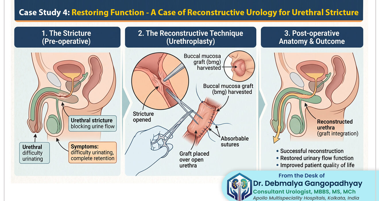
For Rahul, the recommended line of treatment was a Substitution Urethroplasty using a Buccal Mucosa Graft (BMG).
What is a Buccal Mucosa Graft?
“Buccal mucosa” is the medical term for the inner lining of your cheek. But why use cheek tissue for the urethra?
- It is hairless.
- It is accustomed to a wet environment (saliva in the mouth, urine in the urethra).
- It has an excellent blood supply, meaning it “takes” or survives very well when transplanted.
- It is robust and resists shrinking.
By opting for this advanced procedure, Rahul was tapping into the highest standard of Reconstructive Urology Treatments in Kolkata, designed for long-term success rather than a temporary fix.
Preparing for Surgery
Before the surgery, Rahul underwent standard pre-operative blood tests, an ECG, and an evaluation by our anesthesiology team. We instructed him on oral hygiene—using a specialized mouthwash for a few days prior to surgery to ensure the cheek tissue was sterile. He was admitted to Apollo Multispeciality Hospitals the evening before the procedure.
Step-by-Step Breakdown of the Surgery
On the morning of the surgery, Rahul was placed under general anesthesia. The procedure involves two distinct surgical sites working in tandem.
Step 1: Harvesting the Graft (The Mouth)
- While one part of the surgical team prepared the pelvic area, another specialized surgeon worked on the inside of Rahul’s cheek.
- We carefully measured and excised a strip of tissue measuring exactly 5 centimeters long and 1.5 centimeters wide from the inner lining of his cheek.
- The cheek wound was then meticulously closed with dissolvable stitches. (Remarkably, the inside of the mouth heals incredibly fast, usually within a few days).
- The harvested tissue—the graft—was then prepared. We carefully scraped away any underlying fat or muscle to ensure only the pure mucosal lining remained. This thin layer is crucial for absorbing blood and nutrients in its new location.
Step 2: Accessing the Stricture (The Urethra)
- An incision was made in the perineum (the area between the scrotum and the anus).
- We carefully dissected through the tissue layers to expose the bulbar urethra.
- Once located, we opened the urethra lengthwise, cutting exactly through the dense scar tissue, opening it up much like a book. This immediately relieved the physical narrowing.
Step 3: The Reconstruction (Placing the Graft)
- Now came the most crucial part: patching the “pipe.”
- We took the prepared cheek tissue (the BMG) and tailored it to fit the open defect in the urethra perfectly.
- Using incredibly fine sutures (stitches thinner than a human hair), we sewed the cheek graft onto the urethra, widening the tube significantly. We utilized a technique called a dorsal onlay, where the graft is placed against the strong, vascular tissue of the erectile bodies, providing it with excellent blood supply and mechanical support to prevent it from ballooning out in the future.
Step 4: Securing the Healing Process
- Before closing up, we placed a soft silicone urinary catheter through the penis, past the newly grafted area, and into the bladder. This catheter acts like a scaffolding or a stent. It keeps the urethra open, allows urine to bypass the surgical site, and lets the new graft heal undisturbed.
- The perineal incision was then closed in layers.
The surgery took approximately three and a half hours. It was a complex, multi-stage reconstruction executed flawlessly.
Part 5: The Road to Recovery (Post-Operative Care)
When dealing with reconstructive urology, the surgery is only 50% of the battle. The other 50% is how the patient heals.
The First 48 Hours
Rahul woke up in the recovery room. Pain management was a priority. He experienced some soreness in his perineum (surgical site) and a sensation similar to a severe sore throat or mouth ulcer on the inside of his cheek.
- Diet: He was started on a liquid diet (cold soups, ice cream, cold milk) to soothe the cheek, gradually moving to soft foods over the next two days.
- Mobility: By the evening of the surgery, we had Rahul sitting up, and by the next morning, he was taking short walks in the ward. Early mobilization is key to preventing blood clots.
He was discharged on the third post-operative day. The primary challenge he faced at home was managing the urinary catheter.
Living with the Catheter
The silicone catheter had to remain in place for exactly three weeks. For a young, active man, this is psychologically and physically frustrating. We provided Rahul with a leg bag (a smaller urine bag strapped to the thigh under loose trousers) so he could walk around comfortably without carrying a large medical bag in his hand.
We educated him on:
- Keeping the catheter insertion site clean.
- Avoiding heavy lifting, strenuous exercise, or riding motorcycles/bicycles.
- Taking prescribed antibiotics to prevent catheter-associated infections.
- Managing occasional bladder spasms (the bladder’s natural urge to push the catheter balloon out), for which we prescribed specific muscle relaxants.
Though uncomfortable, Rahul understood that this 21-day period was the crucial incubation time for his cheek tissue to permanently integrate into his urinary tract.
Part 6: Follow-Up and The Moment of Truth
The most anticipated day in a urethroplasty patient’s journey is the day the catheter comes out.
The 3-Week Mark: Catheter Removal
Rahul returned to the clinic exactly 21 days post-surgery. Before simply pulling the catheter out, we needed to ensure the internal wound had sealed perfectly. We performed a Pericatheter Urethrogram.
- We injected a small amount of dye alongside the catheter.
- The X-ray showed a beautifully wide, watertight urethra with absolutely no dye leaking outside the grafted area. The graft had taken successfully.
The catheter was gently removed.
Then came the moment of truth. We asked Rahul to drink a few glasses of water and wait until his bladder was full. When he finally went to the restroom to urinate naturally for the first time in three weeks, the result was life-changing.
He came out of the restroom with tears of relief in his eyes. The flow was powerful, effortless, and fast. There was no straining, no waiting, and no pain. A post-removal Uroflowmetry test confirmed a peak flow rate of 28 milliliters per second—a massive jump from his pre-surgery 4 ml/sec. He was completely voiding like a normal, healthy 28-year-old.
The 3-Month and 6-Month Check-ups
Success in reconstructive urology isn’t just about the first day; it is about long-term durability.
- At 3 months: Rahul reported zero issues. His mouth had healed so perfectly that he couldn’t even feel where the tissue was taken from. His urinary flow remained strong.
- At 6 months: We performed a follow-up Uroflowmetry and a routine ultrasound. The bladder wall thickening had started to reverse, proving that the removal of the obstruction was allowing his bladder to heal.
Part 7: Success and Restored Quality of Life
The success of Rahul’s reconstructive repair goes far beyond medical metrics and flow charts. It is about the restoration of human dignity and quality of life.
Before the surgery, Rahul was a prisoner to his bladder. He avoided long road trips, felt anxious during office meetings, and suffered quietly out of embarrassment.
Today, six months post-surgery, Rahul is back to his vibrant self. He sleeps soundly through the night without waking up to use the washroom. He performs his job effectively without the looming dread of bathroom breaks. Most importantly, the psychological weight of a chronic, worsening illness has been lifted off his shoulders.
This case stands as a testament to the fact that urethral strictures, no matter how complex or long-segment they are, can be definitively cured with the right surgical expertise.
Frequently Asked Questions (FAQs) on Reconstructive Urology
To further demystify this topic, here are answers to some of the most common questions patients ask when diagnosed with a urethral stricture.
Can a urethral stricture be cured with medicines?
No. A urethral stricture is a physical, anatomical barrier made of dense fibrous scar tissue. Medicines cannot dissolve this scar. While medications might be given to treat associated infections or relax the bladder temporarily, surgery is the only definitive treatment to remove or bypass the blockage.
Why do urologists take tissue from the mouth (cheek) and not somewhere else?
The inner cheek lining (buccal mucosa) is the “gold standard” for urethral reconstruction. It is naturally hairless, highly flexible, has a rich blood supply ensuring good healing, and is biologically adapted to being in a moist environment (saliva), which translates perfectly to the moist environment of the urethra (urine). Taking skin from the arm or leg often leads to hair growth inside the urethra or shrinkage of the graft over time.
Does taking tissue from the mouth affect eating or speaking?
No, not in the long term. The mouth is one of the fastest-healing areas of the human body. Patients may experience soreness, similar to a bad mouth ulcer, for about 5 to 7 days. You will be on a soft diet for a few days, but within two weeks, most patients report completely normal eating, drinking, and speaking without any lasting pain or visible external scars.
Will the stricture come back after Urethroplasty?
Buccal Mucosa Graft (BMG) Urethroplasty has one of the highest success rates in urology, typically ranging between 85% to 90% for long-term cure. While there is always a minimal risk of recurrence, it is vastly superior to older methods like simple dilatation or laser cutting (VIU), which have very high recurrence rates for long strictures.
How long will I need to stay in the hospital?
For a standard buccal mucosa graft urethroplasty, patients are usually admitted for 3 to 4 days. You can walk the day after surgery, but you will go home with a urinary catheter that stays in place for about three weeks to protect the newly grafted tissue while it heals.
Will this surgery affect my sexual function or fertility?
Urethroplasty is performed on the urinary pipe (urethra), not the prostate or testicles. The vast majority of patients experience no negative impact on erectile function or fertility. In fact, by relieving the pain and psychological stress associated with severe urinary obstruction, many patients report an improvement in their overall sexual health and well-being post-recovery.
Is a catheter painful to live with for three weeks?
It is more frustrating than painful. You will feel its presence, and occasionally, you might experience a bladder spasm (a sudden urge to pee despite the catheter). Your doctor will prescribe medication to minimize this discomfort. It requires care and hygiene, but millions of patients manage it successfully at home every year.
When can I return to normal activities and work?
If you have a desk job (like software development, writing, or administration), you can often return to remote/work-from-home duties within 10 to 14 days, even with the catheter in place. However, heavy physical labor, gym workouts, running, or riding a two-wheeler must be strictly avoided for at least 4 to 6 weeks after the surgery to prevent trauma to the healing perineum.
What happens if a urethral stricture is left untreated?
Ignoring a stricture is incredibly dangerous. The bladder has to push much harder to force urine through the narrow gap. Over time, the bladder muscle thickens and eventually weakens, losing its ability to contract. This backward pressure can push infected urine up into the kidneys, leading to severe kidney damage, recurrent severe urinary tract infections (UTIs), and life-threatening conditions like acute urinary retention (complete inability to pass urine).
How do I know if I need a Reconstructive Urologist?
If you have a weak urine stream, take a long time to urinate, feel incomplete emptying, or have been diagnosed with a stricture that has failed previous simple treatments (like dilatation), you need a specialist. Reconstructive urology is a highly specialized niche within the urology field that focuses entirely on these complex rebuilds.
Why Choose the Right Expert?
The urethra is a delicate structure. Every time a surgical intervention fails, the resulting scar tissue makes the subsequent surgeries much more difficult and lowers the chances of a successful outcome.
This is why, if you are diagnosed with a stricture, seeking out a dedicated Reconstructive Urology Specialist Doctor in Kolkata for your first definitive surgery is vital. The precision required to harvest the graft, prepare the tissue bed, and suture it seamlessly under magnification requires years of specialized training and experience.
If you are experiencing symptoms like a weak urinary stream, frequent nighttime urination, or burning sensations, do not wait for an emergency. Early diagnosis makes the reconstructive process smoother and the recovery faster.
Your quality of life is not something you should have to compromise on. Modern reconstructive urology holds the key to restoring function, confidence, and peace of mind.
Read Also:
- Case Study 1: Overcoming Complex Kidney Stones-A Successful RIRS Procedure
- Case Study 2- Life After Prostate Cancer: Successful Uro-Oncology Management
- Case Study 3: Minimally Invasive Relief: Treating Advanced BPH with Laparoscopic Surgery
Disclaimer:
The information provided in this case study is for educational and informational purposes only and does not constitute medical advice. Patient details have been modified to ensure strict confidentiality and privacy. Every patient’s medical condition, anatomy, and response to treatment are unique. The surgical outcomes described here represent a specific clinical scenario and may not guarantee identical results for all individuals. If you are experiencing urinary symptoms, it is imperative to consult a qualified urologist for a personalized diagnosis and treatment plan. Do not disregard professional medical advice or delay seeking it based on the contents of this article.
For appointments and consultations regarding: Reconstructive Urology Treatments in Kolkata, please visit our clinic or contact us via our official portal. Follow for more updates on Youtube & Facebook